This Is Your Brain on fMRI
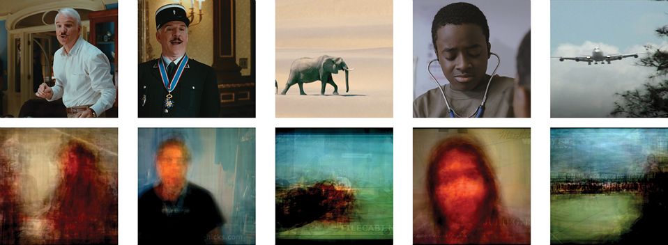
Neuroscientists at the University of California, Berkeley, have used functional magnetic resonance imaging (fMRI) to produce rough representations of images as actually seen by a subject’s visual cortex.
First, researchers created computer models of fMRI signals from each subject’s visual cortex while the subject watched Hollywood movie trailers. Then the scientists recorded visual cortex activity during a second set of clips. They then simulated what the brain “saw” during the second set by running their models on some 5000 hours of online video clips to isolate snippets of images and motions that most closely matched the visual cortex’s activity in each moment.
That said, no one expects cutting-edge scientific laboratories, let alone consumer technology, to be able to decode the brain’s activities in any way that could enable actual mind reading. It’s more that scientists have learned the first few phrases in a very complex language. Worse, the phrases rarely have a fixed meaning and don’t combine to form sentences in a straightforward way.
Geraint Rees, director of the Institute of Cognitive Neuroscience at University College London, has been looking for the equivalent of a Rosetta Stone. “Is looking at red plus drinking a glass of wine equivalent to the sum of looking at red and, separately, drinking a glass of wine? Or is there some kind of nonlinear interaction? We don’t know the rules of those combinations.”
Moreover, says Chris Frith, emeritus professor at the Wellcome Trust Centre for Neuroimaging at University College London, no one has figured out how to read an individual neuron without opening up a skull and attaching a tiny wire to it. Instead, he says, fMRI reads the activity of clusters of hundreds of thousands of neurons. And even the best electroencephalogram (EEG) technology still can resolve only down to the level of hundreds of neurons.
Source


0 Comments:
Post a Comment
<< Home